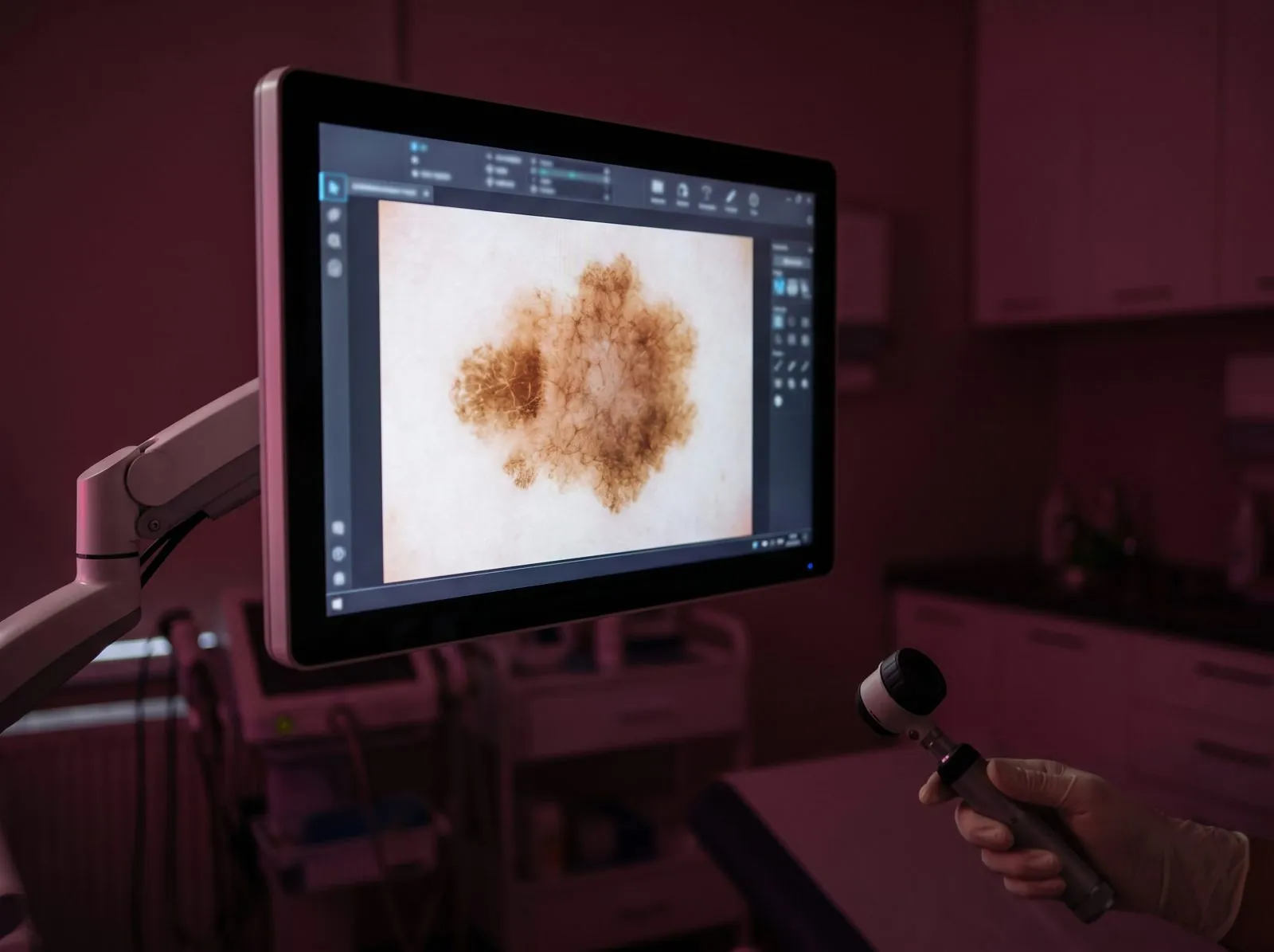

It is used to map the melanocytic lesions of the skin, in order to monitor and prevent the onset of skin tumours, and makes it possible to follow over time the evolution of a skin lesion that you do not want to remove. Video documentation of naevi is essential for the early recognition of skin cancer. These "mole maps" give a panoramic view of all the moles. In the next phase all the naevi are digitally marked and recorded dermatoscopically. The microphotos are automatically linked to the markings. With regular follow-up visits you will be able to see the changes at a glance!
FotoFinder technology:
- Early recognition of changes through follow-up checks of the naevi
- Fewer excisions of harmless moles and improved diagnostic safety
- The full range of diagnosis, from direct-light microscopy to fluorescence diagnosis
- Documentation of treatments and conditions with excellent panoramic photos, from psoriasis to parasitic infestations